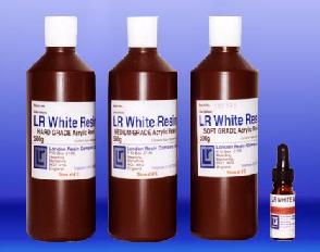
 EM(電顕)及びLM(光顕)用 LRホワイトレジン EM(電顕)及びLM(光顕)用 LRホワイトレジン
(1) LRホワイトレジン EM(電顕)、LM(光顕)
EM(電顕)及びLM(光顕)用二重目的の新しいアクリル樹脂です。 極低粘性で浸透に要する時間が短い上、三通りの硬度が得られる寿命の長いのが特徴です。 特に免疫染色時、電子顕微鏡用, 光学顕微鏡用にご使用下さい。 Accelelatorを使用して低温重合或いは熱重合を選択する事が出来ます。
※2025年3月より販売形態が変更になりました。 これまでは主剤にCatalyst(触媒)が混合された状態で販売しており冷蔵(4℃)保管が必要でしたが、今後は混合せずセットに同梱して販売します。 そのため冷蔵保管の必要がありませんが、ご使用前にユーザー様ご自身で触媒を主剤に混合させる必要があります。 (混合後は冷蔵保管が必要になります。
LRホワイトレジンキット セット内容:
LRホワイトレジン(Medium・Hard・Softいずれか) 500ml 1本 触媒 1本 アクセラレータ 10ml 1本
室温保管可能ですが、ご使用前に触媒を主剤に混合して下さい。 触媒混合後は冷蔵(4℃)保管が必要です。 ゼラチンカプセルで重合して下さい。
The uncatalysed version of LR resin does not need to be kept in a fridge and can be kept at room temperature in a dry place. 24 hours prior to use, simply add in the catalyst(9.9g of catalyst per 500ml of LR White), mix thoroughly and leave for 24 hours to ensure the catalyst has fully dissolved into the resin.
 |
#06-1001 LRホワイトレジンMediumキット |
500g+10ml促進剤 | 88,000.-円 |
| #06-1002 LRホワイトレジンHardキット |
500g+10ml促進剤 | 88,000.-円 |
|
| #06-1003 LRホワイトレジンSoftキット |
500g+10ml促進剤 | 88,000.-円 |
※ 各LM作成用促進剤10ml付き
LR White: A convenient and economical premixed resin with very wide application. Being both hydrophilic and electron beam stable it is equally suitable for light and electron microscopy, and with appropriate fixation the same specimen may be used for both techniques. Published work shows that immunocrytochemical methods may be used through LR White sections without etching or any pre-treatment.
Using LR White for Electron Microscopy (Micrograph: Human Oral Epithelium, P.T.A. stained)
When using LR White embedding resin for dedicated electron microscopy, very few changes need to be made to the regime used for epoxy resin embedding. Every laboratory has its own individual embedding schedule but we have laid out a Typical Schedule for LR White as guidance for its use included to the Kits.
Using LR White for Light Microscopy (Light Micrograph: Human Lymphoma. Hematoxylin & Eosin stained. 2micron section.)
Resin embedding for light microscopy provides greatly improved cellular definition compared to paraffin embedding, and for this reason is now widely used in diagnoses particularly of Renal disease, Lymphomas and bone marrow trephines, as well as research.
The acrylic resins currently used however are not suitable for E.M. and the eooxy resins used for E.M. are not easily stained for light microscopy. LR White, however, can be used for both purposes and lymph node for example (12x10x3mm) can be processed, cut and stained for light microscopy, the same block trimmed down, cut and staied for electron microscopy.
LR White can also be used for the histochemical demonstration of some of the more resistant enzymes, and for the immunocytochemical demonstration of intracellular immunoglobulins.
For those laboratories already using an acrylic ressin e.g. HEMA or Glycol Methacrylate no alteration need be made to the current processing schedule, but we have laid out a typical schedule for LR White as guidance for its use included to the Kits.
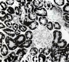
Using LR White for Electron Microscopic Immunocytochemistry Immuno-Gold stained Kidney(Micrograph courtesy of Dr.D.Kerjaschki,Vienna University)
L.R. White resin has five advantages which can be exploited for the localisation of antigens in sections of fixed and embedded tissue under the electron microscope.
It is a hydrophilic embedding agent which means that ultrathin sections allow the passage of aqueous solutions even at neutral pH as opposed to epoxides and polyesters, ultra-thin sections of which are much less permeable.
Its lipid solvency is apparently low for a plastic embedding agent and therefore membrane and cytosol structures can be observed under the electron microscope even when osmium has not been used to stablise lipids. No low temperature methods are required although tissue structure is much improved by perfustion fixation methods.
It does not, where it has so far been tested, prevent the demonstration of antigens by immunochemical techniques. No etching or protease digestion has so far been necessary.
It is beam-stable, standing up well to even quite low KVs, thus representing a considerable advance over the more commonly used methacrylates.
It tolerates rapid, partial dehydration, accepting tissue from 70% ethanol. Such tissue has an improved antigenicity over tissue which has been fully dehydrated.
Using LR White for Immunohistochemistry Picture:Human Lymph Node stained for lg G showing positively staining Plasma Cessl. PAP Method.
Sections from L.R. White embedded tissue have been used sucessfully for immunocytochemistry at both the light microscope and electron microsope levels. This demonstrates quite clearly that the visualizetion steps of the immunocytochemical procedure will penetrate the resin and react with tissue antigens if they have been preserved in the tissue.
Much interest has centered on using immunocytochemistry to detect protein hormones and the various classes of immunoglobulin and generally these classes of antigen have proved resistant to alteration both in processing to paraffin-wax an to L.R. White resin.
It is the special hydrophilic nature of L.R. White which allows immunochemicals to permeate the supporting resin and reach its sites of binding and no resin pertreatement is necessary, or indeed possible, to faciliate this penetration.
We have been sucessful with L.R. White blocks only when they have been thermally cured, probably because when accelerator-curing the resin the exotherm produced is sufficient to damage the integrity of the tissue antigen. Some workers have also reported that a slight under-cure of L.R. White, say at 50° for 20-24hr aids subsequent penetration of antisera, but we have obtained good results without deviating from the standard polymerisation schedule. The guidance included.
Using LR White for Hard Tissue (Picture: Bone from Human Mandible)
LR White can be used for the microtomy of decalcified bone and teeth and also for microtomy or sawing and grinding of undecalcified tissues.
Decalcified Tissue: May be processed, cut and stained similarly to soft tissue (see Using LR White for Light Microscopy), except that dehydration and infiltration times may need to be extended depending on the size of tissue. It is also recommended that bone be de-fatted to imporove the penetration of resin into marrow cavities. This can be be achieved by using chloroform after dehydration, returing to absolute alchohol to remove the chloroform before infiltrating with resin and polymerising.
Undecalcified Tissue: Dehydration and infiltration times will vary depending on size and density of tissue. Those laboratories using Methyl or Butyl methacrylate at present can use similar dehydration times, but infiltration will probably be shortened due to the low viscosity of the resin.
※ 保存は必ず4℃冷暗所にてお願い致します。
LR White Resin (EM, LM) の一般的な包埋方法
固定: 最終ブロックがEM目的の場合は通常の固定法を採ります。 LM染色の適切な微細構造と広い範囲を必要とするならば、フレッシュなパラホルムアルデヒド(3〜4%)を 2½% W/V蔗糖と共にリン酸緩衝液 PH7.2 において使用することが適切です。
脱水: 濃度別したエタノールシリーズを選択します。
浸透: 極度に低い粘性ですので、浸透性が速く、大きな試料にも利用する事が出来ます。 1mm立法の動物細胞試料では室温でレジン、4〜6回、約3時間。
重合: EM レジンの表面は酸素との接触を避け60℃±2の温度で20〜24時間。 LM 冷却保存処理ではレジン10mlに対してアクセルレーター1滴の割合で使用します。 10分〜20分で重合が完了します。 その他、レジンの量とアクセルレーターの量で重合時間を調節する事が出来ます。 紫外線重合にはアクセルレーター0.5µをレジン10mlの割合で加えると、低温重合が可能です。 24〜48時間。
トリミングと切削: 一般的な切削スピードは1秒あたり1mm位が適当です。
染色: 一般的な染色が可能で良い結果が得られます。
※ 個々の使用目的には添付されている Using LR White を参照して下さい。
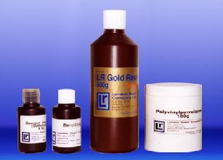
(2) LRゴールドレジン (LR Gold Resin)
新しいタイプのアクリル樹脂で一般的な組織化学法によって、無固定の組織を低温で浸透させ、反応しやすい酵素類保存を良くする。 また親水性なので反応中の基質と抗体の通行を妨げず、正確な位置づけを確実にします。 Semi-thin Ultrathin Section の染色は半永久的であり電子線にも安定しています。 -25℃低温紫外線重合。
#06-1004 LRゴールドResin Kit 価格お問合せ下さい。
内容 LR Gold Resin 500gx1 Benzil 50gx1 Polyvinyl Pyrrolidone 100gx1 LR Gold: A Special acrylic resin for very special purposes. Its infiltration and polymerisation at low tempperatures down to−20℃ means that unfixed tissue may be embedded in LR Gold. This enables enzyme histochemistry and immunocytochemistry of many fixation sensitive enzymes and epitomes to be performed on 1〜2µm resin setions. Bringing the quality of resin histology to an area where only cryostat setions were previously available. LR Gold is a real step forward in histochemial technique. This resin has the ability to be cured by blue light thus making expensive ultra-violet light sources required by other systems unnecessary.
| 〒101-0021東京都千代田区外神田5-3-11 |
| 応研商事株式会社 |
| TEL:03(5826)8642 FAX:03(5826)8643 |
| URL: http://www.okenshoji.co.jp |